Matted Lymph Nodes Ultrasound

Pdf Ultrasonography Of Superficial Lymph Nodes Benign Vs Malignant

Pin By Dr Abuaiad On Lymphatics With Images Sonogram

Tuberculous Lymphadenopathy Radiology Case Radiopaedia Org

Ultrasonographic Differentiation Of Benign From Malignant Neck Lymphadenopathy In Thyroid Cancer Kuna 2006 Journal Of Ultrasound In Medicine Wiley Online Library
Reactive Vs Malignant Lymph Nodes Ultrasound Features Radiology Reference Article Radiopaedia Org

Sonography Of Abdomen Showing Matted Peripancreatic Lymph Nodes Download Scientific Diagram
Level i constitutes lymph nodes above the anterior and posterior bellies of the digastric muscle cephalad to the hyoid bone and inferior to the inferior border of the mandible and includes the submental group of nodes.
Matted lymph nodes ultrasound. In gynecological oncology the peripheral lymph nodes that are especially interesting from a clinical point of view are the inguinal axillary and scalene lymph nodes figure s42 among which particular attention is paid to the inguinal lymph nodes on ultrasound examination as these are regional nodes for vulval vaginal and ovarian cancer. What might cause this answered by dr. So i ve been referred to an oncology surgeon. For levels ii iv the posterior border is the posterior edge of the sternomastoid muscle and the anterior border is the laryngeal complex.
My doctor is concerned because it s a lymph node and matted. The lymph nodes that become inflamed are in a membrane that attaches the intestine to the abdominal wall. Mesenteric lymphadenitis is an inflammation of lymph nodes. Most axillary nodes are reactive or inflammatory someti.
Ultrasound says it looks benign and not enlarged. Cervical adenitis refers to the inflammation of lymph nodes in the neck. These lymph nodes are among. Clinical presentation in the pediatric population a chil.
Transverse view of the abdomen in the region of epigastrium shows hypo echoic conglomerate masses adjacent to aorta and at the level of porta hepatis suggestive of enlarged matted para aortic and porta hepatis lymph nodes giving the appearance of a mass.

Ultrasound Of Malignant Cervical Lymph Nodes Abstract Europe Pmc
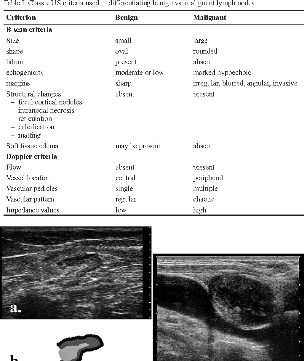
Table I From Ultrasonography Of Superficial Lymph Nodes Benign Vs Malignant Semantic Scholar
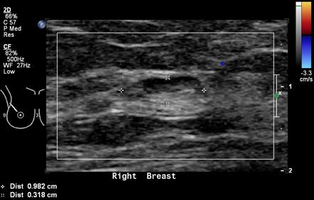
Intramammary Lymph Nodes Radiology Reference Article Radiopaedia Org

Pin By Stelios Daskalogiannis On Digestive In 2020
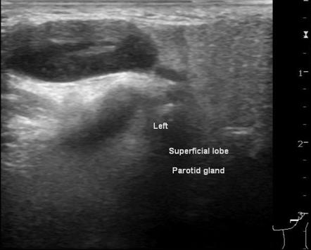
Intraparotid Lymph Nodes Radiology Reference Article Radiopaedia Org

Periportal Lymphadenopathy In Hepatitis A Radiology Case Radiopaedia Org

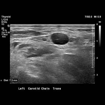
Benign Lymph Node The Normal Neck Contains Scores Of Lymph Nodes Some Of Which Are Easily Seen With Ultrasound This L Ultrasound Thyroid Ultrasound Radiology

Abdominal Lymphoma Radiology Case Radiopaedia Org

Pdf Ultrasound Of Malignant Cervical Lymph Nodes

Pdf Mistakes In Ultrasound Diagnosis Of Superficial Lymph Nodes

Normal Lung Ultrasound With A Lines Ultrasound Lunges Youtube

View Image

Abdomen And Retroperitoneum 1 7 Peritoneum Mesentery And Omentum Case 1 7 2 Mesenteric Lymph Nodes Ultrasound Cases

