Maternal Floor Infarction Radiology
Ethan Frome
Ultrasound Evaluation Of The Placenta And Umbilical Cord Radiology Key
Https Www Radnet Com Hudson Valley Radiology Sites Hvr Files Radnet Imce Maternal Fetal Maternal Fetal Provider Case 20studies 20in 20obstetrical 20mr Pdf

Infarction Infarction Placenta
Https Www Radnet Com Hudson Valley Radiology Sites Hvr Files Radnet Imce Maternal Fetal Maternal Fetal Provider Nj Mfm Society 9 18 14 Pdf
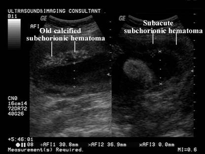
Subamniotic Hemorrhage

It commonly increases with gestational age.
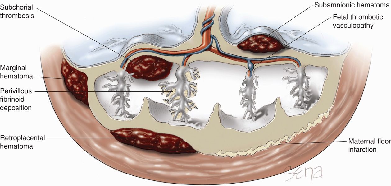
Maternal floor infarction radiology. Am j obstet gynecol 1990 163 935 8. 15 my doctor called with the results of my clotting disorder tests and luke s autopsy clotting disorders such as a factor v or factor ii mutation are one cause of stillbirths. Placental calcification has been considered a manifestation of aging of the placenta. Maternal floor infarction mfi is a poorly understood placental lesion reportedly associated with intrauterine growth restriction iugr and recurrence.
Histological definitions association with intrauterine fetal growth restriction and risk of recurrence. Katzman pj genest dr. Maternal floor infarction frequently recurs in successive pregnancies rate 39 2 and there is evidence that it develops rapidly. In this study of mfi and the related placental disorder massive perivillous fibrin deposition mfd semiquantitative histologic criteria for these diagnoses are defined and rates of iugr and.
Maternal floor infarction and or massive perivillous fibrin can cause stillbirth and can recur. Maternal floor infarction is a relatively rare condition characterized clinically by severe early onset fetal growth restriction with features of uteroplacental insufficiency. The association of maternal floor infarction of the placenta with adverse perinatal outcome. When volume of infarcts is over 50 of placental volume think of lupus anticoagulant.
Maternal floor infarction and massive perivillous fibrin deposition. Pathological characteristics include massive and diffuse fibrin deposition along the. Maternal floor infarction and massive perivillous fibrin deposition. Maternal floor infarction posted on october 26 2015 by luke s mom on thursday oct.
Histological definitions association with intrauterine fetal growth restriction and risk or recurrence. The fibrin extends into the intervillous space where it envelops the. It has a very high recurrence rate and carries a significant risk or fetal demise. It is a disorder characterized by heavy deposition of fibrin in the region of the basal villi immediately adjacent to the decidua basalis.
Delayed placental calcification maternal diabetes rh sensitization accelerated placental calcification norma. Often the lesion is associated with excessive x cell proliferation and cyst formation figure 275. And such are most often associated with pre eclampsia particularly the hellp syndrome variety hemolysis elevated lfts low platelets with rbc fragments. Placenta maternal floor infarction.

Placental Surface Cysts Detected On Sonography Brown 2002 Journal Of Ultrasound In Medicine Wiley Online Library

Infarction Placental Infarcts
Https Www Pedrad Org Linkclick Aspx Fileticket Lteikkg Otu 3d Portalid 5

Myocardial Calcification In A Fetus Mitra 2004 Journal Of Ultrasound In Medicine Wiley Online Library
Fetal Growth And Well Being Fetal Therapy
Placental Multiple Chorionic Cysts In Maternal Scleroderma Springerlink
Placental Abnormalities Obgyn Key

Imaging And Assessment Of Placental Function Moran 2011 Journal Of Clinical Ultrasound Wiley Online Library

Classification Of Placental Lesions American Journal Of Obstetrics Gynecology
Https Onlinelibrary Wiley Com Doi Pdf 10 7863 Jum 2004 23 10 1385
Https Onlinelibrary Wiley Com Doi Pdf 10 1002 Jcu 1870180315

Ultrasound Part Ii Clinical Emergency Radiology
Mri Of The Neonatal Brain Mary A Rutherford

