Mat With Av Block Ecg
First Degree Av Block Av Block I Av Block 1 Ecg Echo

First Second Type 1 And Type 2 And Third Degree Heart Blocks

First Degree Atrioventricular Av Block Ecg Review Criteria And Examples Learntheheart Com

Ecg Case 1 Question 3 Answer Learntheheart Com

Understanding Atrioventricular Blocks Careercert
Multifocal Atrial Tachycardia Mat Litfl Ecg Library Diagnosis

Electrocardiographic effects of first degree av block.
Mat with av block ecg. They are caused by an alteration in the atrioventricular node or in the bundle of his although they can also be caused by malfunctions in. First second and third degree av block may all be diagnosed using the ecg. Management and treatment of av block 1 2 and 3 is discussed in a separate article. Grauer is the sole proprietor of kg ekg press and publisher of an ecg pocket brain book.
Normally the heart rate is controlled by a cluster of cells called the sinoatrial node sa node. Second degree av block mobitz type ii. Two p waves between the qrs complexes are evident while one is hidden over the t wave. Click here for a larger image.
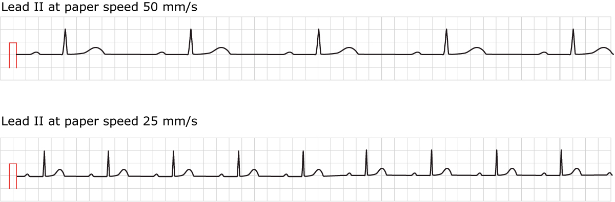
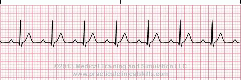
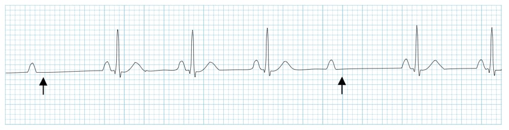
The normal pr interval in an. Therefore the effects of first degree av block are going to be seen in the pr interval namely that the pr interval will be longer than normal just a reminder. Av block can be described by degree based on ecg appearance or by anatomic level of block. The block is located in the atrioventricular node in most cases.
First degree av block is rarely serious and may be left untreated in the majority of cases. First degree av block second degree av block complete av lock. Hence this is a 3 1 av block. Treatment of second degree av block mobitz type 1.
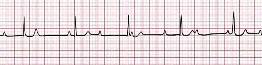
Ecg quiz 55 3 1 av block. Pr interval 0 22 s. At an initial look it may be called 2 1 av block because there are only two obvious p waves between any pair of. Av block 1 av block i 1st degree av block the term block is somewhat misleading in this case because first degree av block only implies that the conduction is abnormally slow.
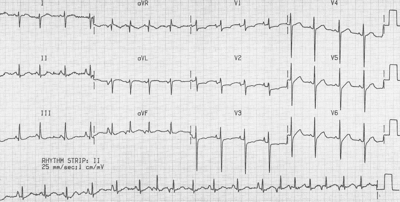
The pr interval is constant although it may be prolonged. The goals of therapy are to treat symptoms and to prevent syncope and sudden card. Ecg criteria for first degree av block. Thinking back to the normal ecg the av nodal delay is the primary determinant of the pr interval.
All p waves are followed by qrs complexes. First degree av block synonyms. Multifocal or multiform atrial tachycardia mat is an abnormal heart rhythm specifically a type of supraventricular tachycardia that is particularly common in older people and is associated with exacerbations of chronic obstructive pulmonary disease copd. Atrioventricular blocks are a set of disorders of the cardiac conduction system that cause the atrial electrical stimulus to the ventricles to be delayed or interrupted 1.
The degree of av block or anatomic level of block does not necessarily correlate with the severity of subsequent symptoms. Av block or blocked pacs. Mobitz type 2 block implies that some atrial impulses are blocked sporadically. The pr interval is 0 22 s in first degree av block.
Atrioventricular Av Heart Block Cardiovascular Medbullets Step 2 3
Different Types Of Heart Block From New To Icu
Av Block 2nd Degree Mobitz Ii Hay Block Litfl
Complete Heart Block Ecg Weekly
Multifocal Or Multiform Atrial Tachycardia Mat Is An Abnormal Heart Rhythm Specifically

Ecg Interpretation Of Arrhythmias Tusom Pharmwiki

Second Degree Heart Block Type I Ekg Answer Page

3rd Degree Av Block Ecg 2 Learntheheart Com

Wide Qrs Complex With First Degree Av Block Ecg Guru Instructor Resources

Second Degree Av Block Mobitz Type 1 Wenckebach Mobitz Type 2 Block Ecg Echo

Conduction Abnormalities
Overview Of Atrioventricular Av Blocks Ecg Echo

Ecg Heart Blocks Ecg Educator Blog Heart Blocks Heart Blocks Ekg Interpretation Nursing School Notes

